New immune cells discovered in the Alzheimer’s brain
Researchers at the Institute of Anatomy at Leipzig University, in collaboration with international partners, have identified a previously unknown subgroup of immune cells in the brain tissue of Alzheimer’s patients. The discovery was made possible by a newly developed microscopy technology that has been specially optimized for the human brain. The results were published in the journal “Nature Neuroscience”.
The newly discovered cells are specialized microglial cells, the brain’s defense cells. This cell population occurs much more frequently in the Alzheimer’s brain and is closely spatially connected with protein deposits typical of the disease. The study also shows that microglial cells can take on various specialized states in Alzheimer’s disease.
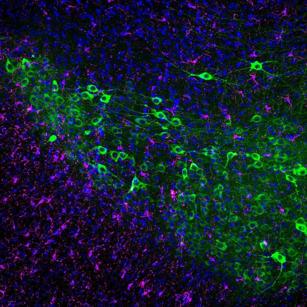
The researchers led by Dr. Dennis-Dominik Rosmus further developed the so-called CODEX method and adapted it for use in the human central nervous system (CODEX-CNS). For the first time, the method enables a high-resolution, spatial analysis of numerous protein markers simultaneously in the tissue. This makes it possible to characterize complex cell populations in their natural environment ŌĆō a step that was often not possible with conventional methods. The results were also confirmed by classical immunofluorescence staining on brain slices from the Leipzig brain bank.
Microglial cells have long been the focus of Alzheimer’s research, as they play an important role in inflammatory processes and the management of pathological protein deposits. While various subgroups were already known in animal models, comparable detailed studies on the human brain have been lacking so far. The new study closes this gap and provides concrete evidence of disease-specific changes in the immune response in the human brain.
In the long term, the discovery could contribute to a better understanding of the biological mechanisms of Alzheimer’s disease. A more detailed knowledge of how microglial cells react to deposits and cell damage is considered a promising starting point for the development of targeted therapies. The research group plans to further develop the CODEX-CNS method and apply it to other neurological diseases. Future studies will investigate whether the identified cell populations can also be detected in living humans, for example with imaging techniques.
The international cooperation took place with the research group of Prof. Dr. Bahareh Ajami at the Oregon Health and Science University in Portland, USA, among others. The study underlines the importance of studies on human tissue in order to realistically map age- and disease-related processes.
Original Paper:
Read Also:
People with Down syndrome have over 90 percent risk of Alzheimer’s disease – MedLabPortal
Editor: X-Press Journalistenb├╝ro GbR
Gender Notice. The personal designations used in this text always refer equally to female, male and diverse persons. Double/triple naming and gendered designations are used for better readability ected.
Most read
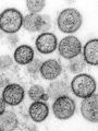
 Hantavirus: detection, symptoms, therapy What is hantavirus? Hantaviruses are a group of viruses that occur worldwide and are mainly transmit...
- Impending fuel shortage: ŌĆ£If the laboratory fails, medical care will come to a standstill in large partsŌĆØ DGKL CEO Jan Wolter explains the dramatic consequences of a potential fuel shortage for laboratory m...
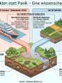
- Hantavirus: Facts instead of panic Reports of three deaths in connection with a hantavirus outbreak on the cruise ship "MV Hondius" in...