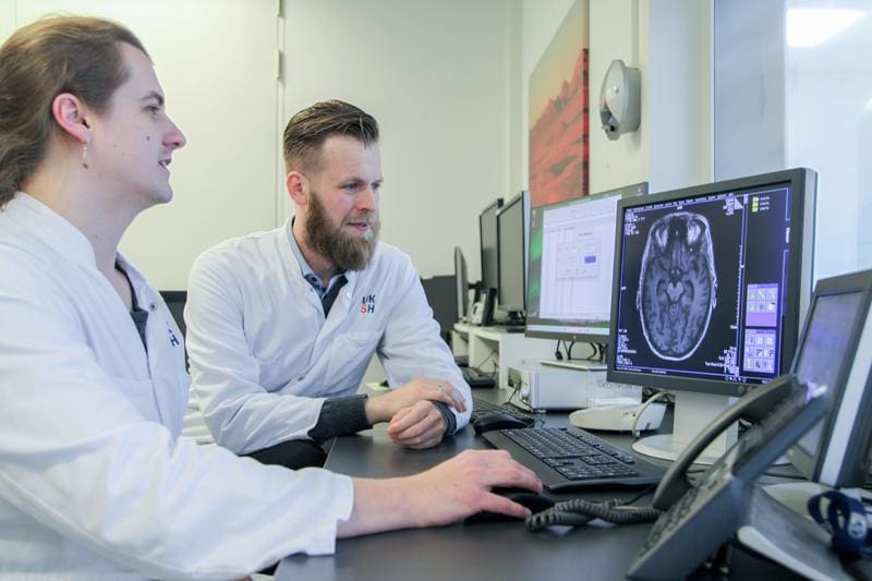
New MRI methods for the early detection of Parkinson’s disease: Campus L├╝beck is looking for study participants
Researchers at the University Medical Center Schleswig-Holstein (UKSH) and the Universities of L├╝beck and Kiel are developing new magnetic resonance imaging (MRI) methods to diagnose Parkinson’s disease earlier and more precisely. The Michael J. Fox Foundation, the world’s largest private sponsor of Parkinson’s research, is supporting the two projects “lysoCEST” and “MiND15” as part of its “Molecular MRI Biomarkers” program with a total of 970,000 US dollars (around 920,000 euros) for the next two years.
Parkinson’s disease is considered the second most common neurodegenerative disease worldwide. In Germany, it is estimated that about 300,000 people are affected, with a further upward trend. The typical movement disorders such as trembling and slowing down are caused by the loss of dopamine-producing nerve cells in the brain. Various cellular mechanisms contribute to the death of these cells ŌĆō but so far there is a lack of clinically applicable methods to detect the individual causes early and non-invasively.
This is exactly where the funded projects come in. The aim is to develop MRI-based imaging methods that make disease-specific biomarkers visible and thus enable earlier diagnosis, more targeted therapy selection and better follow-up. Compared to invasive methods such as cerebrospinal fluid punctures, such examinations would be much gentler on those affected.
In the “lysoCEST” project (funding amount: 570,000 US dollars), PD Dr. Jannik Prasuhn from the Department of Neurology at the UKSH, L├╝beck Campus, is working together with Prof. Dr. Nirbhay N. Yadav from Johns Hopkins University in Baltimore on a method that maps functional disorders of the lysosomes ŌĆō the cellular “waste disposal system”. Such disorders play a central role, especially in patients with GBA1 gene mutations. The researchers want to use an advanced MRI technique to measure and display the accumulation of certain substances as well as changes in the acidity in the lysosomes.
The “MiND15” project (funding amount: 400,000 US dollars) focuses on early disruptions of the mitochondria, the “energy power plants” of the cells. These changes often occur before the first motor symptoms. The team led by PD Dr. Prasuhn and Prof. Dr. Jan Bernd H├Čvener, Head of the Section for Biomedical Imaging at the UKSH, Kiel Campus, and Professor of Translational Magnetic Resonance Imaging at Kiel University, is developing an MRI procedure that is intended to visualize disorders of energy metabolism in the brain with the help of a specially labeled form of vitamin B3 (nicotinamide).
Both approaches will first be tested in the laboratory. If successful, they will be adapted for use in humans and tested in initial clinical trials.
Interested parties can actively support Parkinson’s research at the L├╝beck campus by participating in the “Meta-AdvanceND” clinical trial. Imaging techniques are also used there to investigate the disease biology of neurodegenerative diseases such as Parkinson’s and Alzheimer’s. Further information on participation in the study is available from the Study Center of the Department of Neurology by phone 0451 500-43440 or by e-mail to neuro. MetaAdvanceND@uni-luebeck.de.
Editor: X-Press Journalistenb├╝ro GbR
Gender Notice. The personal designations used in this text always refer equally to female, male and diverse persons. Double/triple naming and gendered designations are used for better readability. ected.
Most read
 Hantavirus: detection, symptoms, therapy What is hantavirus? Hantaviruses are a group of viruses that occur worldwide and are mainly transmit...
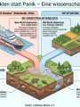
- Hantavirus: Facts instead of panic Reports of three deaths in connection with a hantavirus outbreak on the cruise ship "MV Hondius" in...
- German Medical Association calls for ban on medical diagnostics in drugstores The 130th German Medical Congress 2026 calls on legislators to prevent medical diagnostics in drugst...