Research team optimizes light-sheet microscopy for precise 3D imaging
An interdisciplinary team from the University of G├Čttingen, the University Medical Center G├Čttingen and the University of L├╝beck has developed an innovative platform for light sheet fluorescence microscopes that revolutionizes the imaging of large tissue samples. The technology enables high-resolution, isotropic 3D scans with a resolution of up to 850 nanometers and a speed of 100 frames per second with sample volumes of one cubic centimeter. These advances, achieved through adaptive lighting and optical aberration corrections, open up new possibilities in biomedical research and clinical applications, such as the analysis of neural networks or the planning of surgical procedures.
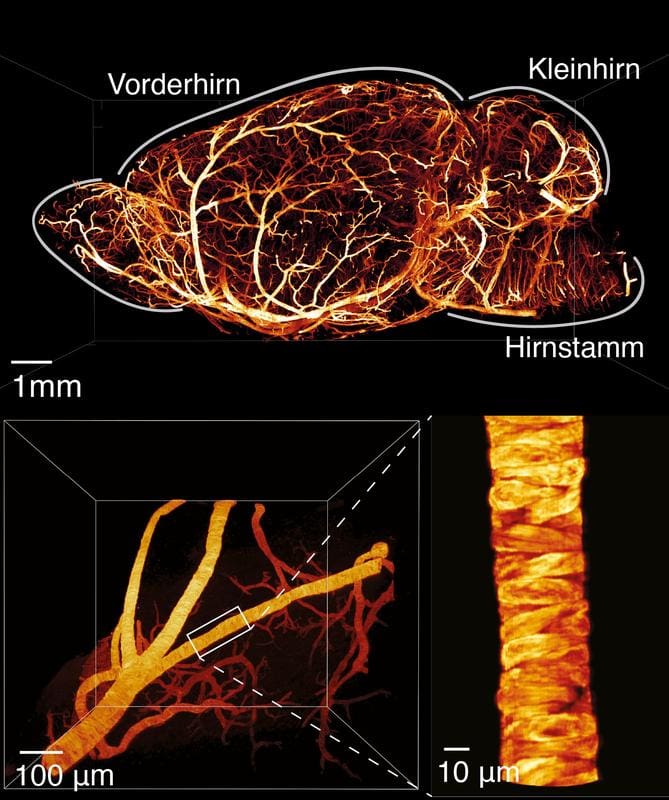
Conventional light-sheet microscopes use a thin layer of light to create layer-by-layer images that are assembled into detailed 3D images. However, they reach their limits in larger chemically transparent tissues: imaging is slow, and optical distortions lead to blurred areas. The new platform overcomes these limitations by combining off-the-shelf components with novel features. As the light sheet travels through the sample, it is continuously readjusted to compensate for refractive index fluctuations caused by clearing methods. Additional algorithms correct aberrations in real time, ensuring uniform sharpness across the entire volume.
The development was carried out as part of the Cluster of Excellence Multiscale Bioimaging, which investigates molecular processes up to cellular networks. The researchers tested the system on various samples, including the cochlea of a mouse, where it mapped fine nerve connections at the single-cell level. Such scans allow insights into healthy and pathological structures, for example in hearing disorders, and could contribute to research into neurodegenerative diseases. Other applications include the visualization of blood vessels or neuronal pathways in entire organs, which advances basic research on cardiovascular systems or brain functions.
The platform is compact, robust and easy to replicate because it is based on accessible components, which makes it easier to distribute in laboratories worldwide. Compared to established systems, it not only doubles the speed, but also improves isotropy ŌĆō the uniform resolution in all spatial directions ŌĆō which is crucial for quantitative analyses. In medicine, it could help with diagnostics by depicting tumor tissue or inflammatory processes in detail, or in surgical planning, where precise 3D models minimize risks.
Original Paper:
Editor: X-Press Journalistenb├╝ro GbR
Gender Notice. The personal designations used in this text always refer equally to female, male and diverse persons. Double/triple naming and gendered designations are used for better readability. ected.
Most read
 Miraculous fitness probiotic: Roseburia inulinivorans massively improves muscle strength Researchers from the University of Granada, the University of Almer├Ła and the Leiden University Medi...
- The personal health check of the UMG laboratory: The detectives of blood The Personal Health Check of the UMG Laboratory (Interdisciplinary Laboratory of the University Medi...
- ASKED: ŌĆ£The new GO├ä is a far greater danger than some people realizeŌĆØ The new fee schedule for doctors (GO├ä) provides for significant cuts for laboratory medicine. DGKL b...