Open-source tool LazySlide facilitates AI analysis of tissue images
A new open-source software tool called LazySlide makes the computer-aided analysis of giant digital tissue images (whole-slide images) much more accessible and easier to combine with molecular data. It was developed by a team led by CeMM researcher Andrûˋ Rendeiro. The study was published on March 20, 2026 in the journal Nature Methods (DOI: 10.1038/s41592-026-03044-7).
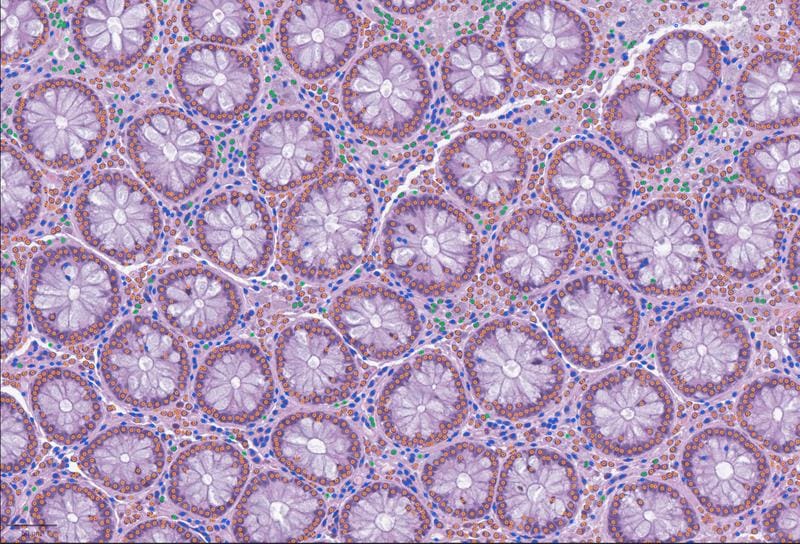
Whole-slide images, which image entire tissue samples in the highest resolution, contain enormous amounts of biological information ã from the overall architecture to individual cells. Despite increasing digitization in pathology and research, this data has so far remained difficult to systematically evaluate and link to other data types such as gene expression profiles. They are often available in proprietary formats and can only be edited with specific, often incompatible programs.
LazySlide uses foundation models ã large AI models pre-trained on extensive data sets ã to break down whole-slide images into smaller, manageable sections and automatically analyze them. The software recognizes tissue patterns, identifies cell types, quantifies structural changes, and connects visual features directly to molecular data without the need for extensive manual annotations.
In an example application, the team examined arterial tissue with and without calcification, a risk factor for cardiovascular disease. LazySlide differentiated between healthy and pathological tissue based on the image features alone and, by integrating RNA sequencing data, made inflammatory signaling pathways visible that were not recognizable in the images alone.
A particular strength of the software is the link with natural language. Researchers can ask specific questions ã for example, where signs of calcification, inflammation or tumor invasion appear in a sample. The AI highlights corresponding regions and provides quantitative assessments. In addition, LazySlide enables zero-shot analyses: For example, the tool recognizes the organ of origin of a sample or distinguishes healthy from diseased tissue without having to be retrained for each question.
The tool is deliberately designed to fit seamlessly into existing bioinformatics workflows in genomics and single-cell research. This is intended to integrate digital pathology more closely into the data-driven ecosystem of the life sciences.
Under the scientific direction of Maria Rescigno, the CeMM ã Research Center for Molecular Medicine of the Austrian Academy of Sciences ã conducts research on cancer, inflammation, metabolic and immune diseases, as well as rare diseases and aging. The facility is located on the campus of the Medical University of Vienna and the General Hospital.
Original Paper:
The study “LazySlide: accessible and interoperable whole slide image analysis” was published in the journal Nature Methods on March 20, 2026, DOI: 10.1038/s41592-026-03044-7
Editor: X-Press Journalistenbû¥ro GbR
Gender Notice. The personal designations used in this text always refer equally to female, male and diverse persons. Double/triple naming and gendered designations are used for better readability. ected.
Most read
 New MRI methods for the early detection of Parkinsonãs disease: Campus Lû¥beck is looking for study participants Researchers at the University Medical Center Schleswig-Holstein (UKSH) and the Universities of Lû¥bec...
- The personal health check of the UMG laboratory: The detectives of blood The Personal Health Check of the UMG Laboratory (Interdisciplinary Laboratory of the University Medi...
- Miraculous fitness probiotic: Roseburia inulinivorans massively improves muscle strength Researchers from the University of Granada, the University of AlmerûÙa and the Leiden University Medi...