Groundbreaking: Dresden team develops modular 3D model of the human liver
Researchers at the Max Planck Institute of Molecular Cell Biology and Genetics have created a three-dimensional organoid model from the patient’s own liver tissue for the first time. The model consists of three cell types ŌĆō adult hepatocytes, cholangiocytes and mesenchymal liver cells ŌĆō and reconstructs the periportal liver region with its structural and functional features.
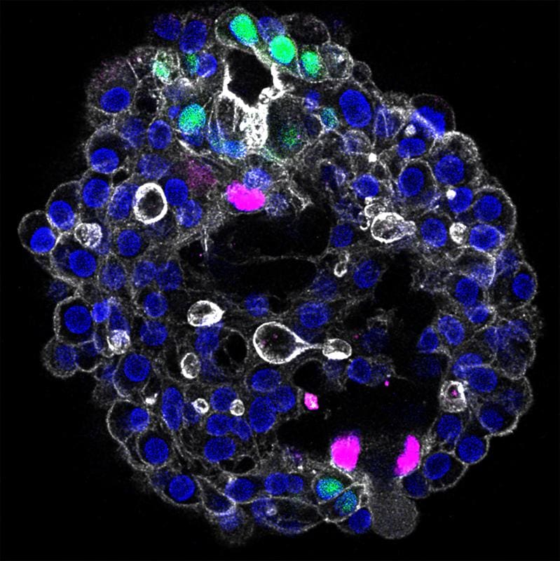
The so-called assembloids imitate liver functions such as detoxification and drug metabolism in the petri dish. Through targeted manipulation, features of bile-induced fibrosis can be replicated, facilitating research into liver disease, accelerating drug development, and promoting personalized approaches.
Previous models were limited to one cell type and could not map the complex cell interaction. The study, published in Nature, is based on collaborations with clinics in Dresden and Leipzig as well as international partners. A biobank with organoids from 28 patients enables further applications in diagnostics and toxicity testing.
Original Paper:
Lei Yuan, Sagarika Dawka, Yohan Kim, Anke Liebert, Fabian Rost, Robert Arnes-Benito, Franziska Baenke, Christina G├Čtz, David Long Hin Tsang, Andrea Schuhmann, Anna Shevchenko, Roberta Rezende de Castro, Seunghee Kim, Aleksandra Sljukic, Anna M. Dowbaj, Andrej Shevchenko, Daniel Seehofer, Dongho Choi, Georg Damm, Daniel E. Stange, Meritxell Huch: Human assembloids recapitulate periportal liver tissue in vitro. Nature, December 17, 2025, doi: 10.1038/s41586-025-09884-1
Editor: X-Press Journalistenb├╝ro GbR
Gender Notice. The personal designations used in this text always refer equally to female, male and diverse persons. Double/triple naming and gendered designations are used for better readability. ected.
Most read
 Miraculous fitness probiotic: Roseburia inulinivorans massively improves muscle strength Researchers from the University of Granada, the University of Almer├Ła and the Leiden University Medi...
- The personal health check of the UMG laboratory: The detectives of blood The Personal Health Check of the UMG Laboratory (Interdisciplinary Laboratory of the University Medi...
- ASKED: ŌĆ£The new GO├ä is a far greater danger than some people realizeŌĆØ The new fee schedule for doctors (GO├ä) provides for significant cuts for laboratory medicine. DGKL b...