Diagnostics: RACOON THREAD revolutionizes early detection of adenomyosis
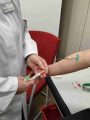
In the RACOON FADEN project, a consortium led by the University Hospital Erlangen and Charit├® – Universit├żtsmedizin Berlin has systematically analyzed MRI images of the uterus in a healthy and diseased state for the first time. The CuraMate software, developed by the Fraunhofer Institute for Digital Medicine MEVIS, was used to annotate, segment and quantify image data in order to develop a method for the early detection of adenomyosis. This disease affects around ten percent of women of childbearing age worldwide and can cause severe pain and infertility.
The focus is on the three-layered uterine wall, consisting of the endometrium, junctional zone and muscle layer. In adenomyosis, endometrial tissue grows into the muscle layer, causing inflammation and dysfunction. The junctional zone serves as a potential biomarker, but its manual determination is too time-consuming for routine clinical practice. CuraMate, a web-based software, enables automated segmentation using deep learning technologies. Radiologists first trace the uterine structures on MRI images in order to train the algorithm, which later recognizes these structures independently. The software, which has been in development since 2017, offers tools for annotation, visualization and workflow optimization, adaptable to different use cases.
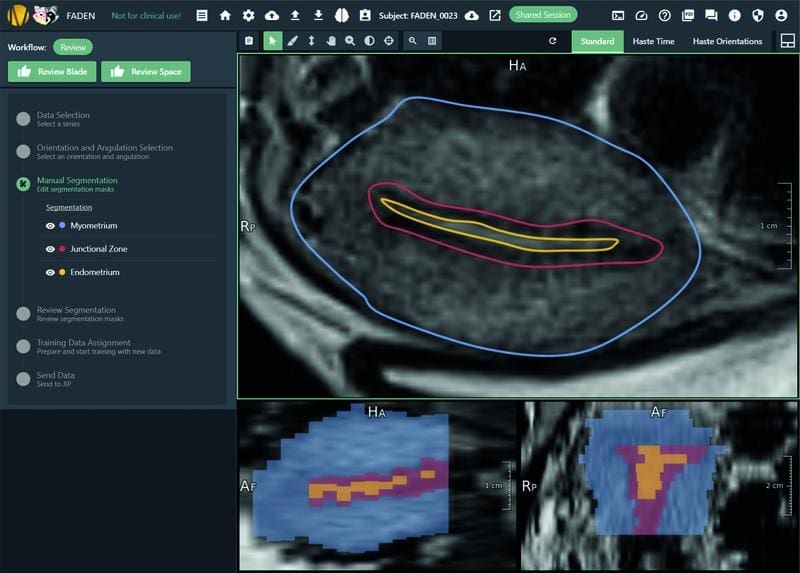
The study was the first to provide MRI reference data for the healthy uterus of women aged between 12 and 30 years. It showed that the uterus takes on different shapes during an MRI examination and does not lie evenly in the body. To take this into account, CuraMate was adapted so that the uterine axis can be aligned before annotation, making segmentations comparable. The junctional zone, previously considered a fixed size, varies depending on the menstrual phase and shows less mobility in women with severe menstrual pain. The wall thickness of 12 millimeters reported in the literature for the early stages of adenomyosis could not be confirmed.
The project, which was funded by the Federal Ministry of Education and Research, ran from January 2024 to June 2025 and involved twelve endometriosis centers in the University Medicine Network. It used the RACOON and NUKLEUS research infrastructures as well as the expertise of the German Cancer Research Center, Mint Medical GmbH and the Helmholtz Zentrum M├╝nchen. The results could provide the basis for more precise early detection of adenomyosis and improve the treatment of affected women.
Editorial office: X-Press Journalistenb├╝ro GbR
Gender note. The personal designations used in this text always refer equally to female, male and diverse persons. Double/triple references and gendered designations are avoided in favor of better readability.
Most read
 New MRI methods for the early detection of ParkinsonŌĆÖs disease: Campus L├╝beck is looking for study participants Researchers at the University Medical Center Schleswig-Holstein (UKSH) and the Universities of L├╝bec...
- Miraculous fitness probiotic: Roseburia inulinivorans massively improves muscle strength Researchers from the University of Granada, the University of Almer├Ła and the Leiden University Medi...
- The personal health check of the UMG laboratory: The detectives of blood The Personal Health Check of the UMG Laboratory (Interdisciplinary Laboratory of the University Medi...