Microfluidics system developed for more reliable super-resolution microscopy of cells
An international research team led by scientists from the University of G├Čttingen and the University Medical Center G├Čttingen (UMG) has presented a novel microfluidic system that significantly simplifies multiplex super-resolution microscopy, makes it more reproducible and gentler on sensitive cell samples. The method enables high-resolution imaging of complex cellular structures and molecular interactions far beyond the limits of conventional light microscopy and is expected to be accessible to a wider range of researchers in the future.
The work was carried out as part of the G├Čttingen Cluster of Excellence “Multiscale Bioimaging: From Molecular Machines to Networks of Excitable Cells” (MBExC) and was published in the journal ACS Nano . The system automates the precise replacement of solutions in the sample chamber, eliminating the need for manual marking and washing steps with pipettes. As a result, conditions remain stable over long imaging cycles, minimizing artifacts and enabling precise mapping of signals from different biomolecules.
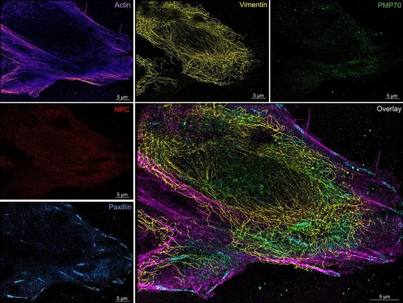
Due to the constant environmental conditions, we can reliably image proteins, specialized cell structures and their complex interactions, explains Dr. Samrat Basak, biophysicist at the University of G├Čttingen (currently LMU Munich). The system proved to be particularly advantageous for sensitive samples: The organization of protein filaments was visualized in human cancer cells. In highly specialized heart muscle cells of a mouse, it prevented deformations or detachment of the cells from the surface ŌĆō a problem that conventional methods often cause.
The microfluidic system can be operated both manually and fully automatically and is compatible with various super-resolution and other imaging methods. It was deliberately designed to be cost-effective and adaptable in order to meet the individual requirements of different biological issues. “We eliminated a central source of error ŌĆō manual fluid exchange ŌĆō and made complex protocols much more user-friendly,” says Dr. Roman Tsukanov from the Department of Multiscale Biology at the University of G├Čttingen, who led the study.
Prof. Dr. J├Črg Enderlein, biophysicist at the University of G├Čttingen and MBExC member, emphasizes the potential for standardization: The approach could make multiplex super-resolution microscopy a widely available method and thus advance both basic research and future medical applications.
Original Paper:
Versatile Microfluidics Platform for Enhanced Multitarget Super-Resolution Microscopy | ACS Nano
Editor: X-Press Journalistenb├╝ro GbR
Gender Notice. The personal designations used in this text always refer equally to female, male and diverse persons. Double/triple naming and gendered designations are used for better readability. ected.
Most read
 German Medical Association calls for ban on medical diagnostics in drugstores The 130th German Medical Congress 2026 calls on legislators to prevent medical diagnostics in drugst...
- Expert Tip for Preanalytics of Glucose Determination The pre-analysis of glucose determination is a science in itself, even for experienced doctors. MedL...
- Preanalytics in glucose measurements: Centrifuged PST tubes are superior to NaF-KOx A study by Brigham and Women's Hospital and Harvard Medical School shows that centrifuged lithium he...