AR holograms to allay fears for parents of children with heart disease
An interdisciplinary team from cardiology, pediatric heart surgery and pediatric cardiology at the University Hospitals of Heidelberg and Mû¥nster is launching the “HoloHeart” project. Augmented reality is to be used to develop a true-to-the-original heart hologram to clearly explain congenital heart defects to parents and children and to reduce fears of surgery. The project is funded by the German Heart Foundation with around 79,000 euros.
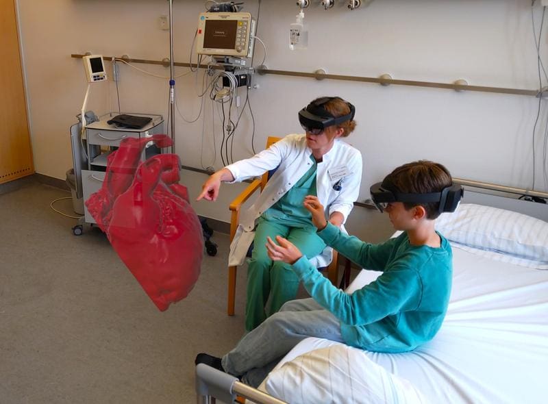
Augmented reality projects three-dimensional organs into space as holograms, visible through special AR glasses. The research team uses this technology to make congenital heart defects in children tangible. In Germany, 8700 children are born with such a defect every year, and more than 95 percent reach adulthood. The project is intended to advance patient-centered medicine by making complex heart defects and treatments understandable and plannable.
The preparatory work began in 2020 with holograms of real hearts for the teaching of medical students. The AR models are created from data from magnetic resonance and computed tomography as well as electrophysiological examinations. They convey complex relationships better than conventional methods. At the same time, virtual 3D models were used on devices or as physical hard sculptures in the clinic, for example for parent education and surgical planning in the case of complicated defects.
A common heart defect is the ventricular septal defect, a hole in the ventricular septum that can lead to overloading. The AR hologram faithfully reproduces the child’s heart and explains the defect and the operation. This takes away parents’ fears. The technology is also suitable for child-friendly explanations: In cooperation with a primary school teacher, content is prepared didactically. Children can touch, turn, enlarge or even go into their heart.
In the case of complex defects such as single-chamber hearts or double outlet right ventricle, AR models offer advantages for planning complicated procedures and increase safety. About one-third of operations in pediatric heart surgery are complex, including re-operations. In pediatric cardiology, holograms help with arrhythmias or catheter interventions, such as ablations, stent or valve implantations.
The project optimises diagnostics and treatment decisions for cardiac malformations. It opens up a new dimension in imaging diagnostics and contributes to anxiety reduction. The Heart Foundation sees this as an innovation boost for pediatric cardiology and cardiac surgery.
The technology could revolutionize reconnaissance and planning. Parents understand the procedure better, which increases the success of the investigation. For surgeons, it improves the visualization of anatomical details. In the long term, AR could be used as standard in pediatric cardiology to improve care and minimize complications. The interdisciplinary team from Heidelberg and Mû¥nster combines expertise in electrophysiology, pediatric heart surgery and pediatric cardiology. The funding enables systematic testing in practice.
Further information:
veldt.ar – Pediatric HoloHeart on Vimeo
Editor: X-Press Journalistenbû¥ro GbR
Gender Notice. The personal designations used in this text always refer equally to female, male and diverse persons. Double/triple naming and gendered designations are used for better readability. ected.
Most read
 Wal Timmy: Environment Minister Backhaus ignored rescue technology that has been in place since 1983 The stranded humpback whale "Timmy" suffered severely for days in Mecklenburg-Western Pomerania beca...
- Wal Timmy: Germanyãs problem with euthanasia EDITORIAL. Letting Timmy suffer is more due to the German lack of experience and the bureaucratic hu...
- Impending fuel shortage: ãIf the laboratory fails, medical care will come to a standstill in large partsã DGKL CEO Jan Wolter explains the dramatic consequences of a potential fuel shortage for laboratory m...