New microscopy method revealed: Cells actively promote flu virus infection
An international research team led by ETH Zurich has used a novel microscopy method to observe live for the first time how influenza viruses infect human cells. The finding: Cells actively support virus uptake. The method could advance the development of targeted antiviral therapies.
Vivid atomic force microscopy, a combination of atomic force and fluorescence microscopy, enables high-resolution real-time observations of the infection process. In contrast to previous methods such as electron microscopy, which destroys cells, or fluorescence microscopy with limited resolution, the new technique shows the dynamics of virus entry in detail.
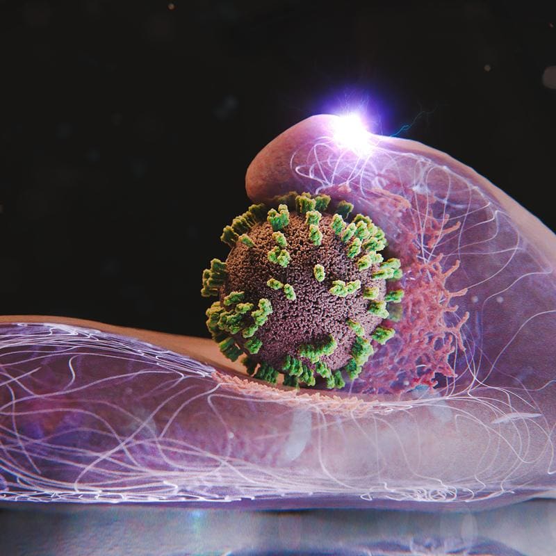
The study reveals that cells are not passively infected by the virus. Influenza viruses use a cellular uptake mechanism that normally transports substances such as hormones or iron. Viruses “surf” on the cell surface, bind to receptor molecules and enter the cell at sites with high receptor density. There, the cell forms a depression through clathrin proteins, which encloses the virus and transports it into the cell interior in a vesicle.
The method shows how cells actively promote this process, for example by recruiting clathrin or wave-like membrane movements that trap the virus. These findings open up new approaches for antiviral drugs, the effect of which can be tested in real time. The technology is also applicable to other viruses and vaccines.
Original Paper:
Yoshida A, Uekusa Y, Suzuki T, Bauer M, Sakai N, Yamauchi Y: Enhanced visualization of influenza A virus entry into living cells using virus-view atomic force microscopy. PNAS, 122: e2500660122, doi: 10.1073/pnas.2500660122
Editor: X-Press Journalistenbû¥ro GbR
Gender Notice. The personal designations used in this text always refer equally to female, male and diverse persons. Double/triple naming and gendered designations are used for better readability. ected.
Most read
 Wal Timmy: Environment Minister Backhaus ignored rescue technology that has been in place since 1983 The stranded humpback whale "Timmy" suffered severely for days in Mecklenburg-Western Pomerania beca...
- Wal Timmy: Germanyãs problem with euthanasia EDITORIAL. Letting Timmy suffer is more due to the German lack of experience and the bureaucratic hu...
- Impending fuel shortage: ãIf the laboratory fails, medical care will come to a standstill in large partsã DGKL CEO Jan Wolter explains the dramatic consequences of a potential fuel shortage for laboratory m...