Cell atlas of the human embryonic brain paves the way for Parkinson’s therapies
Researchers at Duke-NUS Medical School have created the most detailed single-cell atlas of the developing human brain to date. The map includes almost 680,000 cells from fetal tissue, identifies all cell types, their genetic profiles and interactions ŌĆō and sets new standards for the laboratory cultivation of nerve cells. Particularly in focus: dopaminergic neurons of the midbrain, the loss of which triggers Parkinson’s disease. The results, which were recently published in Science Advances (DOI: 10.1126/sciadv.adu7944), provide a reference for developing cell therapies more precisely and excluding unwanted co-cells.
Parkinson’s is the second most common neurodegenerative disease in Singapore; about three out of 1,000 people over 50 are affected. The disease destroys dopamine-producing neurons in the midbrain, causing tremors, stiffness, and loss of movement. “Cell transplantation could alleviate symptoms ŌĆō if the implanted neurons are exactly the same as the human original,” explains Dr. Hilary Toh, MD-PhD candidate in Duke-NOUS’ Neuroscience & Behavioral Disorders program and first author of the study.
BrainSTEM: High-Resolution Two-Step Mapping
The team developed the framework BrainSTEM (Brain Single-cell Two tiEr Mapping). In the first stage, they analyzed almost 680,000 cells from the entire fetal brain with partners ŌĆō including the University of Sydney. The second, high-resolution level zooms into the midbrain and labels dopaminergic neurons with the highest precision. “This reference map allows laboratories around the world to benchmark their cultures against the real human brain,” says Toh.
The analysis showed that many common protocols for growing midbrain neurons also generate cells from other brain regions ŌĆō so-called off-target populations. BrainSTEM detects even subtle contaminants and helps optimize protocols.
Precision for safe cell therapy
“High purity is crucial to minimize side effects and maximize the effectiveness of transplants,” emphasizes Dr. John Ouyang, Principal Research Scientist at Duke-NUS’ Centre for Computational Biology and senior author. The atlas not only provides a blueprint, but also an open-source software package that can analyze other cell types in the brain.
Assistant Professor Alfred Sun, also a senior author, adds: “BrainSTEM sets a new gold standard. It accelerates the development of reliable Parkinson’s models that truly reflect human biology.”
AI and personalized medicine
The high-resolution data will serve as the basis for AI models that are intended to stratify patients and tailor therapies. “We are not only recording cells, but their developmental dynamics ŌĆō a springboard for precise medicine in neurodegenerative diseases,” says Ouyang.
The project was funded by the USyd-NUS Ignition Grant and the Duke-NUS Parkinson’s Research Fund (supported by the Ida C. Morris Falk Foundation). The atlases and the BrainSTEM toolkit are provided as an open-source resource.
Professor Patrick Tan, Senior Vice-Dean for Research: “This work establishes multi-level mapping as indispensable for complex systems. It accelerates research and therapy ŌĆō and gives patients with Parkinson’s new hope.”
Editor: X-Press Journalistenb├╝ro GbR
Gender Notice. The personal designations used in this text always refer equally to female, male and diverse persons. Double/triple naming and gendered designations are used for better readability. ected.
Most read
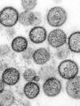
 Hantavirus: detection, symptoms, therapy What is hantavirus? Hantaviruses are a group of viruses that occur worldwide and are mainly transmit...
- Impending fuel shortage: ŌĆ£If the laboratory fails, medical care will come to a standstill in large partsŌĆØ DGKL CEO Jan Wolter explains the dramatic consequences of a potential fuel shortage for laboratory m...
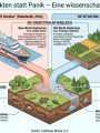
- Hantavirus: Facts instead of panic Reports of three deaths in connection with a hantavirus outbreak on the cruise ship "MV Hondius" in...