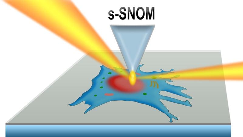
New method enables high-resolution infrared maps of living cells
Infrared vibrational spectroscopy at the synchrotron source BESSY II can be used to create high-resolution maps of molecules in living cells and cell organelles in their natural aqueous environment. This is shown by a study by a team from Helmholtz-Zentrum Berlin and Humboldt-Universit├żt zu Berlin. Nano-IR spectroscopy with infrared-scattering near-field optical microscope is suitable for the examination of tiny biological samples and generates infrared images of molecular vibrations with nanometer resolution. Even three-dimensional information in the form of infrared tomograms is possible. To test the method, the team grew fibroblasts on a highly transparent silicon carbide membrane and examined them in vivo. The method opens up new insights into cell biology.
Infrared microspectroscopy is a non-destructive technique for characterizing biological tissues or cells. With the help of the near-field optical microscope, the smallest sample volumes are sufficient to obtain detailed information about the molecular composition, structure and interactions with a spatial resolution of up to ten nanometers.
The IRIS beamline at BESSY II provides the high-brilliance infrared light required for this method. In the current study, led by Alexander Veber from Helmholtz-Zentrum Berlin and Janina Kneipp from Humboldt University, the team demonstrated the effectiveness of the method by recording vibration spectra of living fibroblast cells in liquids. Fibroblasts are responsible for building connective tissue and producing collagen.
For the first time, the team used an ultra-thin silicon carbide membrane as a biocompatible protective layer between the cells in their liquid medium and the probe tip of the infrared nanoscope, which detects the vibrations.
Not only could the cell nucleus and the cell organelles be visualized, but the contributions of proteins, nucleic acids, carbohydrates and membrane lipids could also be read out on the basis of the recorded vibration spectra. This was possible because the silicon carbide membrane is highly transparent to infrared light. The observed cell structures at the nanoscale correspond to the known heterogeneity of cells and thus confirm the new method.
By varying the measurement parameters, it was possible to control how deep in the sample the signals were detected. In this way, different layers could be examined. This paves the way for infrared nanotomography of cells, i.e. a detailed three-dimensional visualization of cell structure and composition. Standardized two-dimensional and three-dimensional vibrational imaging and spectroscopy could accelerate advances in biophysics and nanomaterials.
The method enables a more precise analysis of biological samples and liquid-solid interfaces than before. In principle, it could be used to study any type of cell, including cancer cells. The development is now available to all user groups of the IRIS beamline.
PREVIEW: The German Congress of Laboratory Medicine (DKLM) 2025 promises exciting insights into the interface between science and clinical practice. Under the motto “Science for Precision Medicine”, the German Society for Clinical Chemistry and Laboratory Medicine (DGKL) and the Umbrella Association for Technologists and Analysts in Medicine Germany (DVTA) invite experts from research, clinics and industry to meet on October 23 and 24 at the Congress Center Leipzig (CCL). The two-day event is aimed at laboratory physicians, biomedical analysts and decision-makers to discuss current advances in diagnostics and strengthen networks. The ceremonial opening of the congress will take place on 22 October with the presentation of the MedLabAwards in the Salles de Pologne.
Editor: X-Press Journalistenb├╝ro GbR
Gender Notice. The personal designations used in this text always refer equally to female, male and diverse persons. Double/triple naming and gendered designations are used for better readability. ected.
Most read
 Wal Timmy: GermanyŌĆÖs problem with euthanasia EDITORIAL. Letting Timmy suffer is more due to the German lack of experience and the bureaucratic hu...
- Rabbit fever infects people in Germany: symptoms, detection and blood count In Lower Saxony and Bremen, three people have already fallen ill with rabbit fever (tularemia) this...
- Blood values in the blue whale (Balaenoptera musculus) Humpback whale Timmy is still keeping Germany in suspense - but what about laboratory values in whal...