New Fraunhofer Method Reduces Radiation Exposure in Breast and Lung Cancer Diagnosis
A new method from three Fraunhofer Institutes combines X-ray and radar technology to make the diagnosis and treatment of breast and lung cancer more precise and less radiation. In the “Multi-Med” project, researchers are developing a multimodal imaging method that significantly reduces radiation exposure compared to conventional computed tomography (CT). While a breast CT scan causes about three times the natural annual radiation exposure of 2.1 millisieverts, the new approach is intended to minimize this risk.

Radar, which has so far been little used in medicine, provides three-dimensional images without health risks and detects tissue changes due to differences in electrical permeability and conductivity. The challenge is to link radar and X-ray data through special co-registration procedures. New reconstruction algorithms improve the image quality of both systems, reduce artifacts in CT and increase the accuracy of detail. Radar data is incorporated into the X-ray reconstruction to reduce radiation exposure.
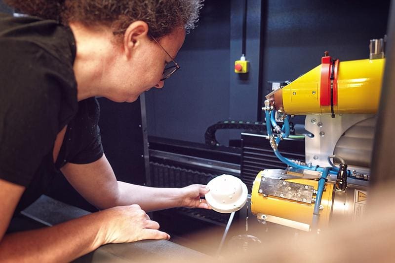
The research team has already developed measurement phantoms that simulate realistic tissue structures to test the method. The aim of the three-year project is a multimodal laboratory system that combines X-ray CT and radar imaging to detect tissue changes early, precisely and with low radiation.
The project is funded by the Fraunhofer-Gesellschaft and led by the Fraunhofer Institute for High-Speed Dynamics, Ernst Mach Institute (EMI), in cooperation with the Fraunhofer Institutes for Digital Medicine MEVIS and for High Frequency Physics and Radar Techniques FHR.
Editor: X-Press Journalistenb├╝ro GbR
Gender Notice. The personal designations used in this text always refer equally to female, male and diverse persons. Double/triple naming and gendered designations are used for better readability. ected.


